Anatomical features of the surface of the eye
anatomical features of the surface of the eye
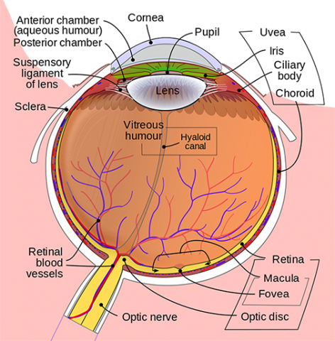
The Eye’s Anatomy
Choroid
Between the retina (the inner light-sensitive layer) and the sclera is a layer of blood vessels that lines the back of the eye (the outer white eye wall).
HEB Scholarship Programme 2023
Auxiliary Body
Muscle-containing structure located behind the iris that focuses the lens.
Cornea
The clear front window of the eye that transmits and focuses light into the eye (i.e., sharpness or clarity). Laser surgery reshapes the cornea, altering the focus.
Fovea
The macula’s centre, which provides sharp vision.
Iris
The coloured part of the eye that helps regulate the amount of light that enters it. When there is a lot of light, the iris closes the pupil to allow less light in. When there is a lack of light, the iris dilates the pupil to allow more light in.
Lens
Light rays are focused onto the retina. The lens is clear and replaceable if necessary. As we age, our lenses deteriorate, necessitating the use of reading glasses. Intraocular lenses are used to replace lenses that have become clouded due to cataracts.
Macula
The area of the retina containing light-sensitive cells. These light-sensitive cells in the macula allow us to see fine details clearly in the centre of our visual field. The deterioration of the macula is a common condition that occurs as we age (age related macular degeneration or ARMD).
The Optic Nerve
A network of over a million nerve fibres that transports visual messages from the retina to the brain. (To see, we need light and our eyes must be connected to the brain.) Because it combines images, your brain actually controls what you see. The retina sees images upside down, but the brain flips them around. This reversal of the images we see is similar to a mirror in a camera. Glaucoma is one of the most common eye conditions caused by damage to the optic nerve.
Pupil
The dark opening in the centre of the iris. The pupil dilates to compensate for the amount of light available (smaller for bright light and larger for low light). This opening and closing of light into the eye is similar to the aperture in most 35 mm cameras, which allows in more or less light depending on the circumstances.
Retina
The nerve layer that runs along the back of the eye. The retina detects light and generates electrical impulses that are transmitted to the brain via the optic nerve.
Sclera
The white coat of the eye that surrounds the iris.
The Vitreous Humor
The clear, gelatinous substance that fills the eye’s central cavity.
How the Eye Functions
Sight, sound, taste, hearing, and touch are the five senses. Sight, like the other senses, is intimately linked to various parts of our anatomy. The eye is linked to the brain and relies on it to interpret what we see.
The transfer of light affects how we see. Light enters the eye through the cornea and travels to the lens. The cornea and lens work together to focus light rays on the back of the eye (retina). The retinal cells absorb light and convert it into electrochemical impulses, which are then transmitted to the brain via the optic nerve.
The eye functions similarly to a camera. A camera’s shutter can close or open depending on the amount of light required to expose the film in the camera’s back. The eye, like the shutter on a camera, works in the same way. The iris and pupil regulate how much light enters the back of the eye. When it is very dark, our pupils dilate, allowing more light to enter. The lens of a camera can focus on objects both far away and close up using mirrors and other mechanical devices. The lens of the eye assists us in focusing but sometimes requires assistance in order to focus clearly. Glasses, contact lenses, and artificial lenses all aid in improving our vision.
This article will teach you about the anatomical features of the eye, including the cornea. The optic nerve is the part of the eye responsible for transmitting electrical signals from the eye to the brain. The eyeball is a sphere with three major components: the retina, pupil, and lens. All of these components work together to provide you with the ability to see. As a result, knowing the correct labels for these structures is critical.
The main part of the retina is the sclera, a tough leather-like tissue that surrounds the eye. The lens and the iris are both made of a single material known as choroid. The choroid layer is the primary source of vision, with the sclera assisting it in focusing. These structures are linked together by muscles known as extraocular muscles. These muscles allow the eye to look up or down and assist the retina in moving in the desired direction.
The retina is a light-sensitive nerve layer located at the back of the eye. When light enters the retina, it is focused onto the retina by the lens, a transparent structure within the eye that focuses light rays. The vitreous humour, a clear jelly-like substance that helps keep the eye round, is found in the centre of the eye. The cornea and sclera are the other two structures.
The cornea is the eye’s clear dome-like surface. The pupil is a thin membrane in front of the eye. The pupil dilates to accommodate changing lighting conditions. The retina is the nerve layer that lines the inside of the eye. It is in charge of interpreting light and transmitting it to the brain. The cornea is the eye’s white outer coating. The vitreous humour fills the central cavity of the eye.
The retina is the light-sensitive nerve layer lining the back of the eye. Because it is the back of the eye, the image it sees is inverted. The retina is a light-sensitive nerve layer located at the back of the eyes. The iris is the clear dome-shaped surface of the eye in front. The sclera is the white coating that surrounds the iris.
The sclera is the tough leather-like tissue that surrounds and shapes the eye. It also houses the extraocular muscles, which allow you to move your eyes up and down. When you are staring at something, you must know the name of the eye. This is the iris, which is located on the outside of the eye. To view the image, you must first label it on the retina.
The sclera is a tough, leather-like tissue that surrounds the eye and gives it shape. There are two chambers in the sclera. The anterior chamber is the inside of the eye, and the posterior chamber is the back of the lens. The anterior chamber receives light and transmits it to the retina. The posterior segment is the eye’s outer coat, while the retina and iris are located on the back surface. The sclera fills the space in the centre of the eye.
The sclera is the tissue that surrounds and shapes the eye. Extraocular muscles attach to the sclera and allow the eye to move to the left and right. They also cause the eye to concentrate. A yellowish-brown sclera lines the sclera. The iris is the first layer of the sclera.
The sclera is the tough leather-like tissue that surrounds and shapes the eye. The extraocular muscles are attached to the sclera and are responsible for moving the eye up and down. When reading or watching television, you can move your gaze up and down, causing the sclera to pull the eye inward. The iris is the coloured part of the eye, and it gets its colour from the macula.
What are the anatomical characteristics of the eye?
The eye is composed of three coats that surround the optically clear aqueous humour, lens, and vitreous body. The cornea and sclera form the outermost coat; the choroid, ciliary body, and iris form the middle coat, which contains the main blood supply to the eye and extends from the back to the front.
What are the seven components of the eyes?
The cornea, iris, pupil, aqueous humour, lens, vitreous humour, retina, and optic nerve are the main components of the human eye.
The Human Eye’s Structure and Function -anatomical features of the surface of the eye
Also read: how to win Morgan Wallen Tickets
What are the eye’s outer parts?
Cornea: The cornea is the clear, outer part of the focusing system of the eye, located at the front of the eye. Iris: The coloured part of the eye that surrounds the pupil is known as the iris. It controls the amount of light entering the eye.anatomical features of the surface of the eye
What are the three layers of the eyeball, and can you describe each one?
The eye is composed of three layers: the outer layer, known as the fibrous tunic, which consists of the sclera and the cornea; the middle layer, known as the vascular tunic, which consists of the iris, choroid, and ciliary body; and the inner layer, known as the…anatomical features of the surface of the eye